
7 Successful management has even been reported with bowel rest and hydration, with no need for antibiotic therapy. Surgical management is extremely rare and antibiotic therapy is the treatment of choice in such cases. Some studies indicate that diverticulitis of the right colon is more benign than that of the left colon. 6 The first 3 characteristics were observed in the CAT scan of our patient, which, added to the intramural air, resulted in the definitive diagnosis. 4 To confirm the diagnosis, computed tomography must show thickening of the colonic wall and an increase in the density or stranding of the pericolonic fat, as well as possible pericolonic abscesses and intramural and extraluminal air. 5 Even less frequent is a clinical picture similar to that of acute cholecystitis secondary to diverticulitis of the right colon or transverse colon, as in our patient, and we know of the report of only one such case.

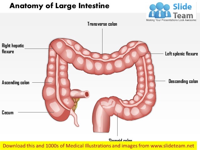
4 The most commonly related presumptive diagnosis in patients with diverticular disease of the right colon is acute appendicitis, with some published case reports. Inflammation or perforation of the diverticula in that zone is very rare, occurring in only 0.5-2.7% of cases. 3 It accounts for only 5 to 10% of all diverticulitis in the large intestine. 2ĭiverticular disease in the transverse colon or hepatic flexure, first described in 1944 by Thompson and Fox, is extremely infrequent. 1 Three main factors are known to cause diverticular disease: disordered intestinal motility, a low-fiber diet, and intestinal wall abnormalities. The present case is a rare variant of diverticular disease, given that the most commonly affected site is the sigmoid colon in white patients and the right colon in Asians. The patient was asymptomatic at one month, then at 12 months, after his original symptoms. Six weeks later, a control CAT scan was performed that showed complete resolution of the perivesicular and pericolonic inflammation. The patient improved and was released under oral treatment with the same antibiotics for another 7 days. Initial management was conservative, with bowel rest, analgesics, and double-regimen antibiotic therapy with ciprofloxacin and metronidazole for 7 days.

Whether the CAT images were caused by colonic pathology or by gallbladder pathology could not be accurately determined. Because the pain continued, a computed axial tomography (CAT) scan with intravenous contrast medium was carried out ( Fig. An upper abdominal ultrasound study identified a distended gallbladder with biliary sludge and a 3 mm wall, a bile duct diameter of 5.5 mm, and the remaining parameters within normal limits. The patient’s hemoglobin level was 12.4 g/dl, platelets 336,000 cells/mm 3, leukocytes 16,400 cells/mm 3, and unaltered liver function tests. Physical examination revealed reduced peristalsis, abdominal distension due to bloating, pain in the right hypochondrium upon palpation, and a positive Murphy’s sign. The pain had no aggravating factors and partially improved with antispasmodics. He did not present with vomiting, fever, or jaundice. It began insidiously, with an intensity of 8/10, and was accompanied by abdominal bloating, hyporexia, and nausea. We present herein the case of a 56-year-old man, with an unremarkable past medical history, who came to the emergency room complaining of abdominal pain of 3-day progression in the right hypochondrium. The journal accepts original articles, scientific letters, review articles, clinical guidelines, consensuses, editorials, letters to the Editors, brief communications, and clinical images in Gastroenterology in Spanish and English for their publication. The scientific works include the areas of Clinical, Endoscopic, Surgical, and Pediatric Gastroenterology, along with related disciplines. The principal aim of the journal is to publish original work in the broad field of Gastroenterology, as well as to provide information on the specialty and related areas that is up-to-date and relevant. Its pages are open to the members of the Association, as well as to all members of the medical community interested in using this forum to publish their articles in accordance with the journal editorial policies. The Revista de Gastroenterología de México (Mexican Journal of Gastroenterology) is the official publication of the Asociación Mexicana de Gastroenterología (Mexican Association of Gastroenterology).


 0 kommentar(er)
